 by
by Cardiac pacing leads play an important role in delivering electrical impulses from implanted cardiac pacemakers to the heart muscles. These thin insulated wires facilitate communication between the pacemaker and the heart and help regulate abnormal heart rhythms.
Types of Pacing Leads
There are mainly two types of pacing leads used – unipolar leads and bipolar leads.
Unipolar leads: A unipolar lead has one electrode at the tip that delivers electrical impulses to the heart and the pacemaker device acts as the other electrode. It is easy to insert but produces a larger electrical field that can stimulate muscles other than the heart.
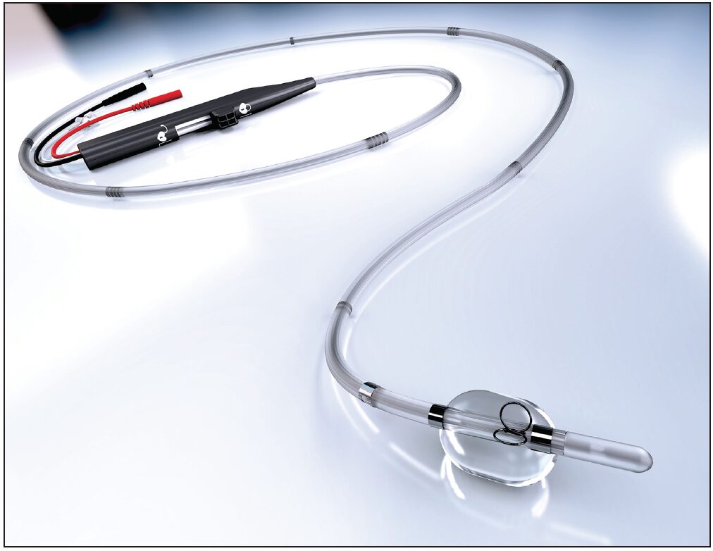
Bipolar leads: A bipolar lead has two electrodes – one at the tip for stimulus delivery and the other, a ring electrode, placed few centimeters above. It targets a specific area and produces a more localized electrical field compared to unipolar leads. However, bipolar leads are technically more complex.
Lead Placement
Cardiac Pacing Leads are placed in the right-sided chambers of the heart – the right atrium or the right ventricle depending on the location that requires pacing.
Right atrial leads: These are usually used for atrial pacing in conditions like atrial fibrillation or sick sinus syndrome. The lead is inserted through a vein in the neck, chest or arm and guided into the right atrium.
Right ventricular leads: For ventricular pacing in heart block or bundle branch block, the lead is inserted through a vein and guided to the right ventricle usually terminating in the right ventricular apex.
Dual-chamber pacing: In some cases, both atrial and ventricular pacing is required which uses separate bipolar leads placed in the right atrium and ventricle respectively. This facilitates synchronized AV pacing and mimics normal sinus rhythm better.
Lead Functions and Testing
The main functions of pacing leads include:
1. Conducting electrical stimuli from the pacemaker to the target chamber of the heart. Modern pacing leads made of alloys like MP35N have high conductivity.
2. Acting as sensors to transmit electrical cardiac signals back to the pacemaker for sensing cardiac activity.
3. Maintaining stable contact with myocardial tissues over years without dislodgment or insulation breaks.
4. Resisting thrombosis or endocardial fibroses at the electrode-tissue interface.
After placement, pacing leads undergo threshold and impedance testing to check their stability and performance. Long-term monitoring helps detect any complications.
Pacing Lead Complications
Although pacing systems have high success rates, certain lead-related complications can occur over time. Some common issues include:
Lead Displacement: Pacing leads can migrate from their original site of placement, especially active young patients. It requires repositioning.
Lead Fractures: Thin insulated wires in the leads are susceptible to fractures, damaging the conduction pathway. It needs lead extraction and replacement.
Insulation Breaches: Cracks in the insulation sheath can short-circuit the lead with cardiac tissues. Recurrent oversensing or failures arise.
Infections: Pacemaker system infections involving the pacing leads are a serious risk, requiring complete system removal.
Thrombosis: Clot formation on the electrode surface hinders proper contact and signal transmission. Anticoagulation medication is used.
Manual Deactivation: Interference from EMI or physical damage can sometimes manually deactivate pacemaker functions that require reprogramming.
Lead Extraction
Infections, broken leads or recalls sometimes necessitate full system extraction along with leads. New sheath technology, locking stylets and powered tools facilitate safer extractions than before with reduced risks of endocardial perforations or cardiac damage. Laser or mechanical powered tools have improved outcomes.
*Note:
1. Source: Coherent Market Insights, Public sources, Desk research
2. We have leveraged AI tools to mine information and compile it